Lumpy skin disease: Clinical signs and postmortem findings
Learn more about the clinical signs of lumpy skin disease 22 June 2023
 1 minute read
 By:
By: Part of Series:
Editors note: The following content is an excerpt from Lumpy Skin Disease: a field manual for veterinarians which is designed to enhance awareness of lumpy skin disease and to provide guidance on early detection and diagnosis for private and official veterinary professionals (in the field and in slaughterhouses), veterinary paraprofessionals and laboratory diagnosticians.
The incubation period in experimentally infected animals varies between four and seven days, but in naturally infected animals it may be up to five weeks.
Clinical signs include:
• Lachrymation and nasal discharge – usually observed first.
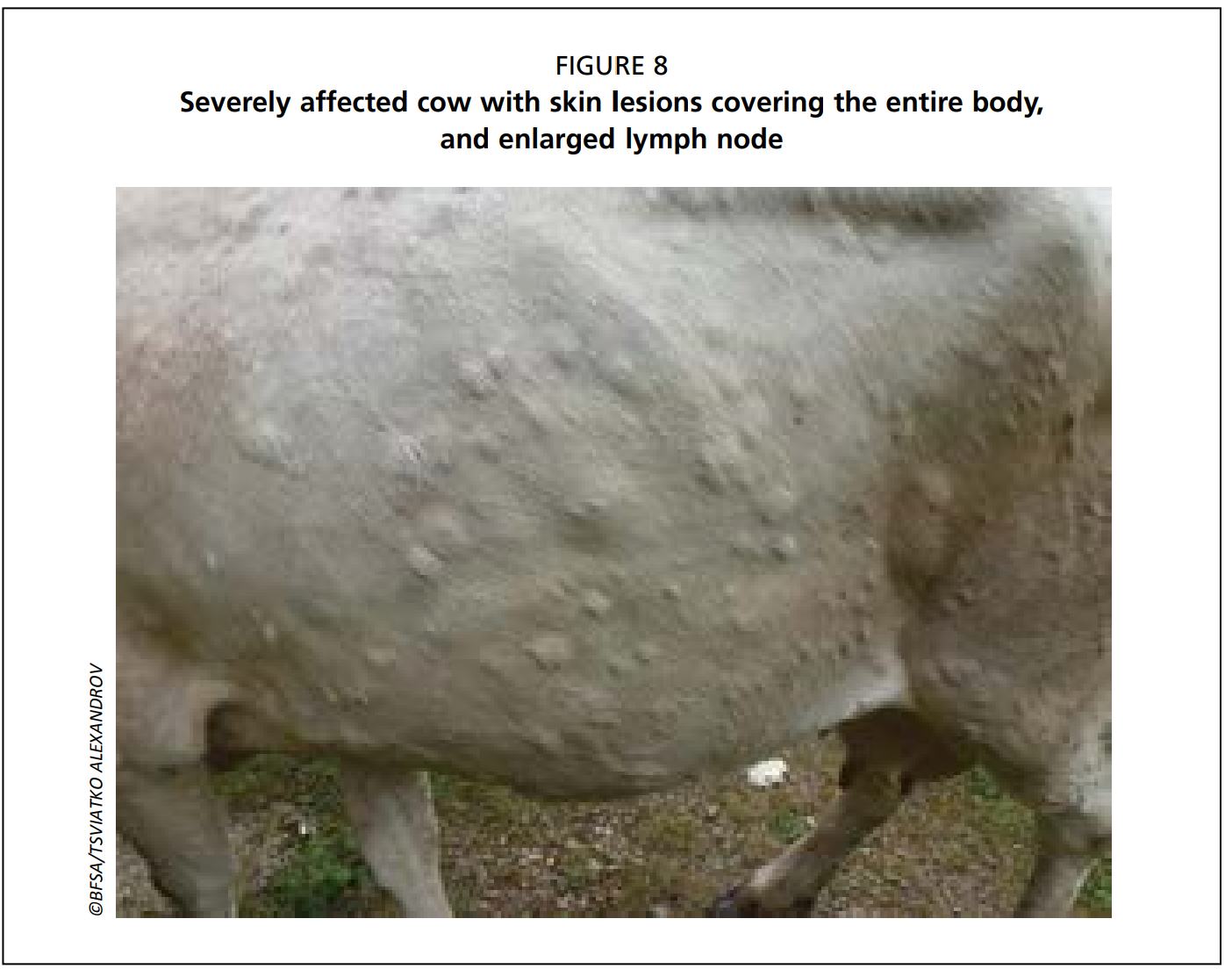
• Subscapular and prefemoral lymph nodes become enlarged and are easily palpable.
• High fever (>40.50C) may persist for approximately a week.
• Sharp drop in milk yield.
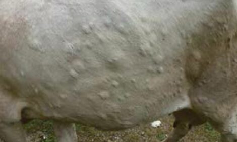
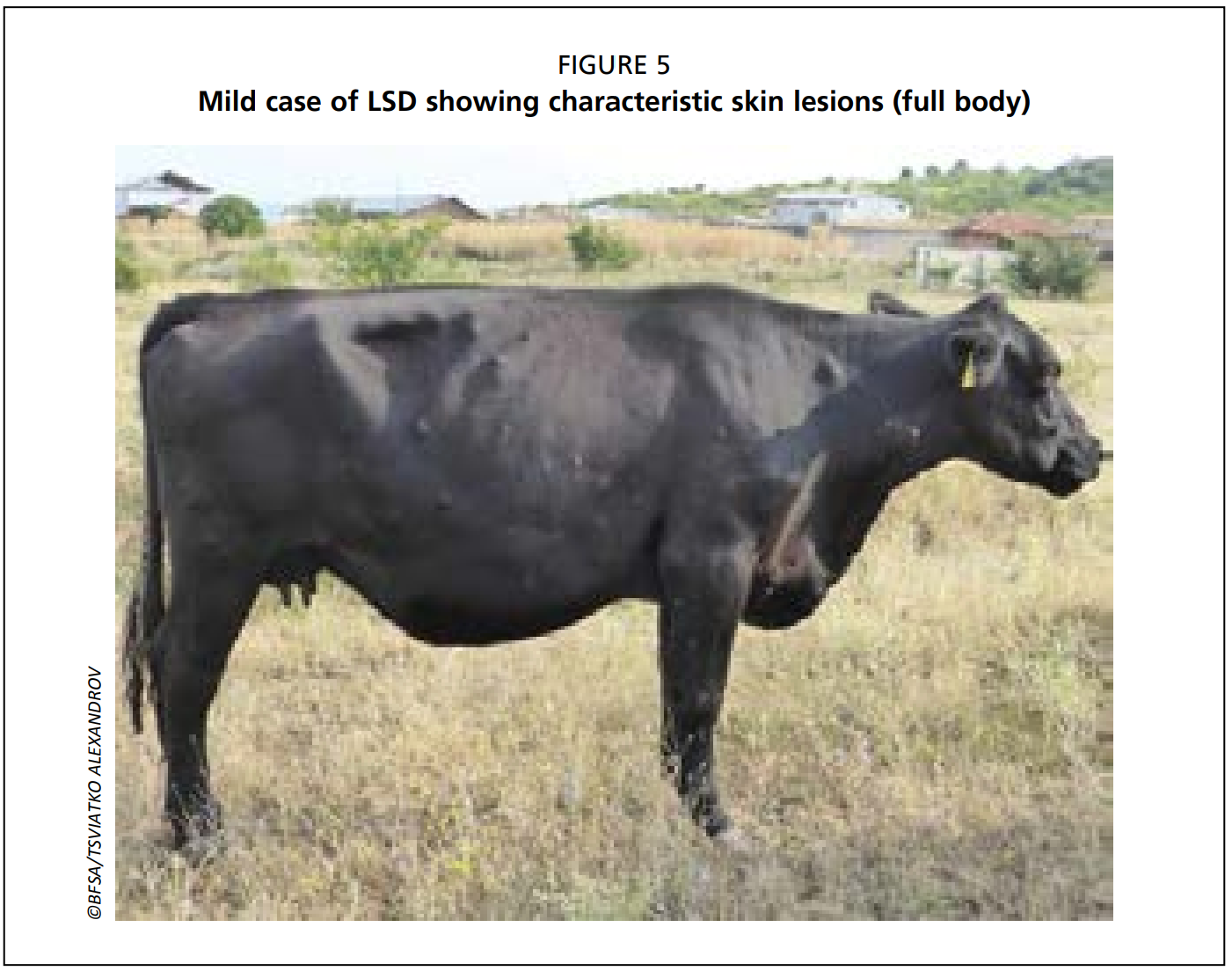
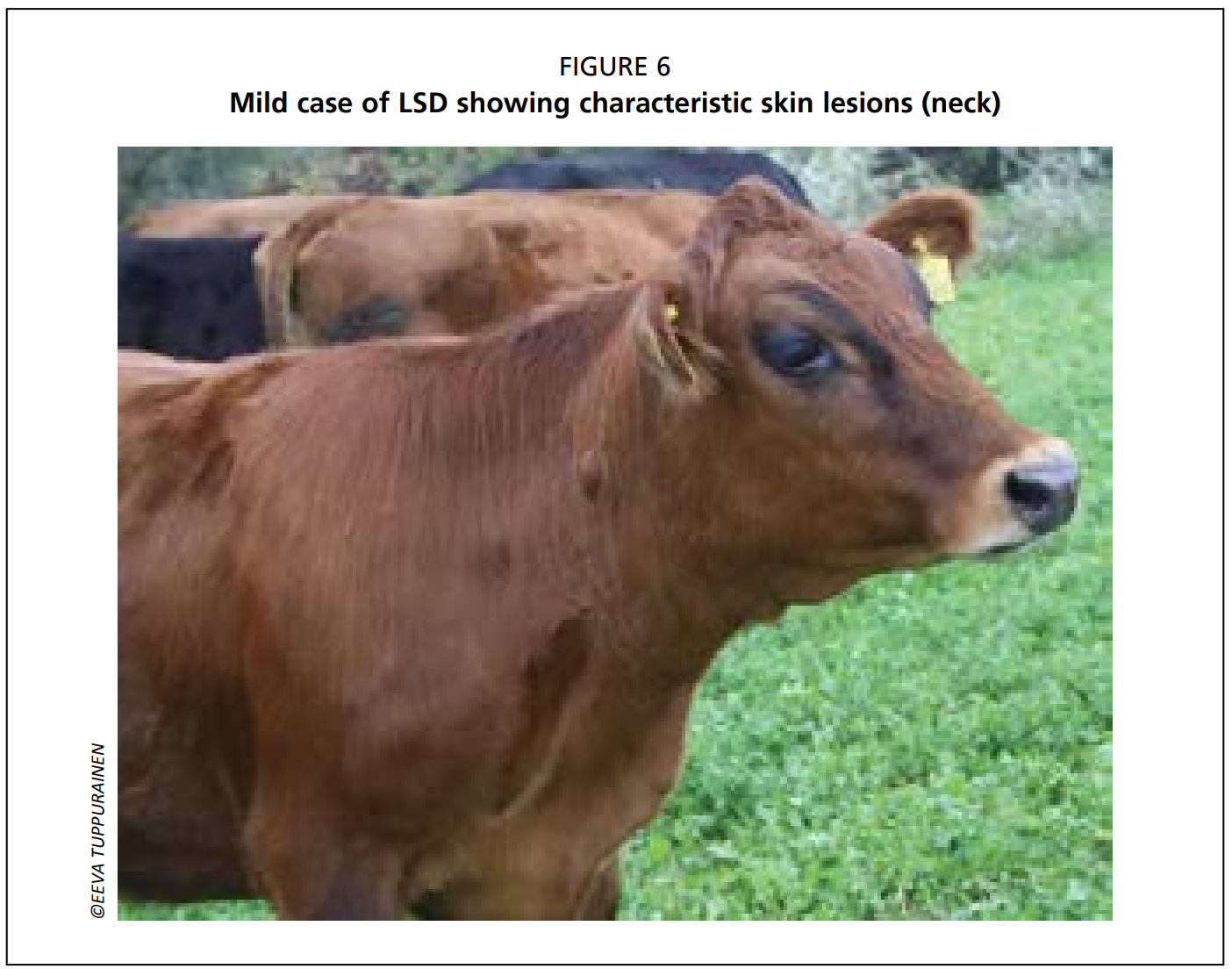
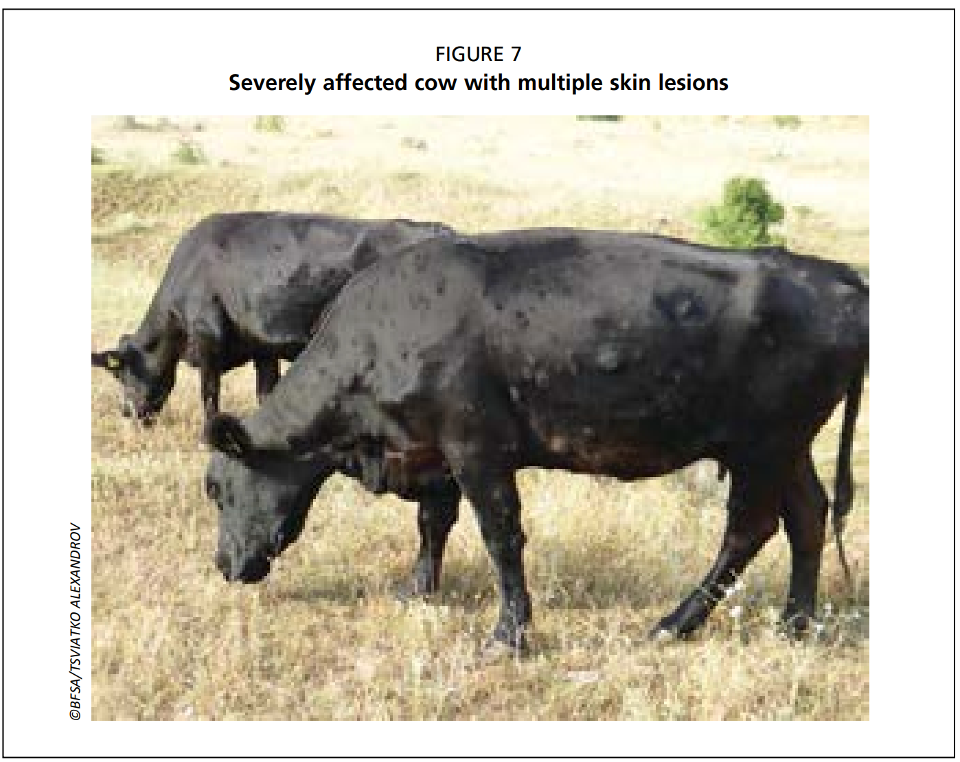
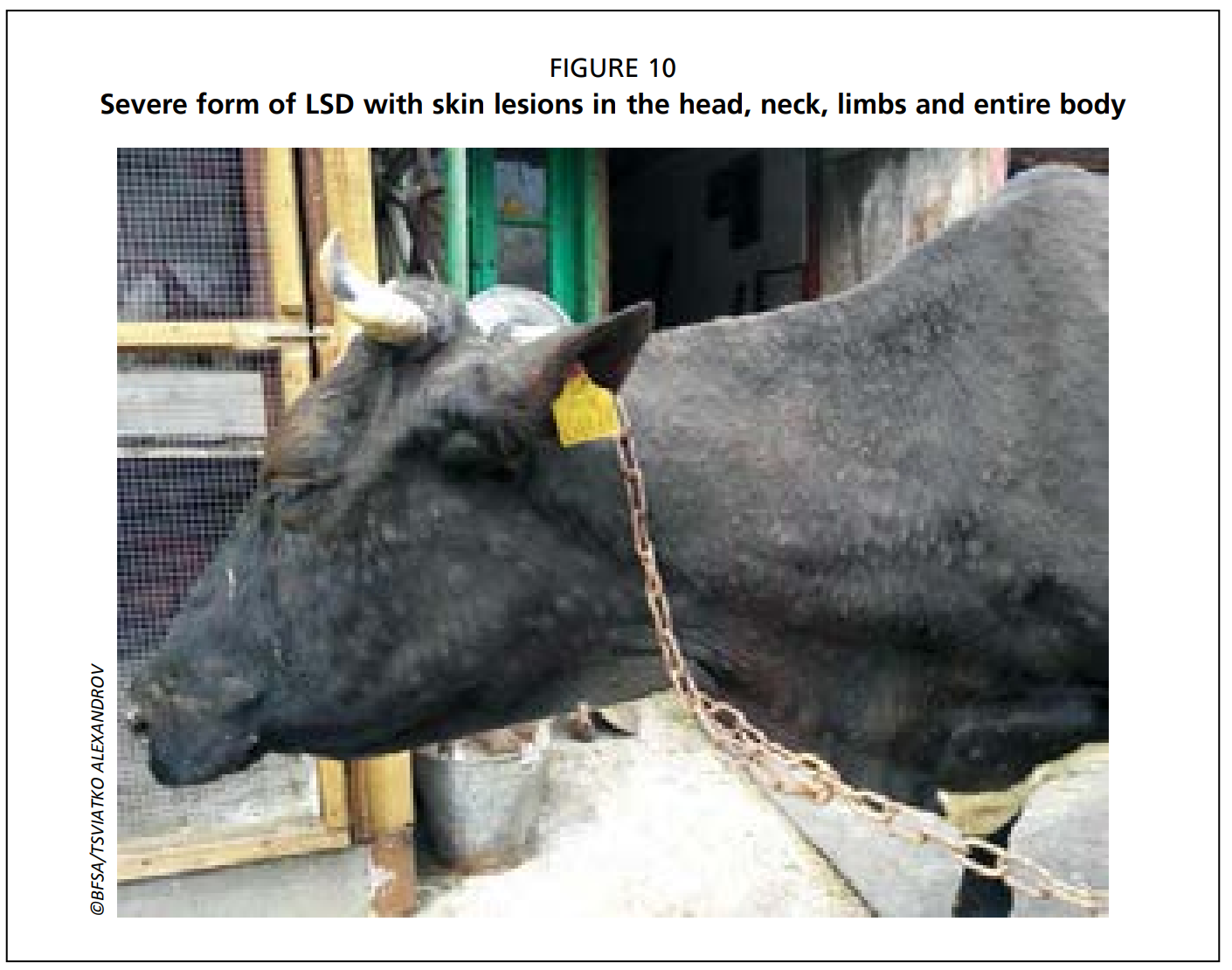
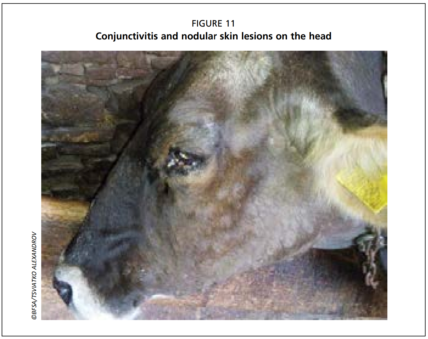
• Appearance of highly characteristic, nodular skin lesions of 10-50 mm in diameter:
- The number of lesions varies from a few in mild cases (Figs. 5 and 6), to multiple lesions in severely infected animals (Figs. 7-10).
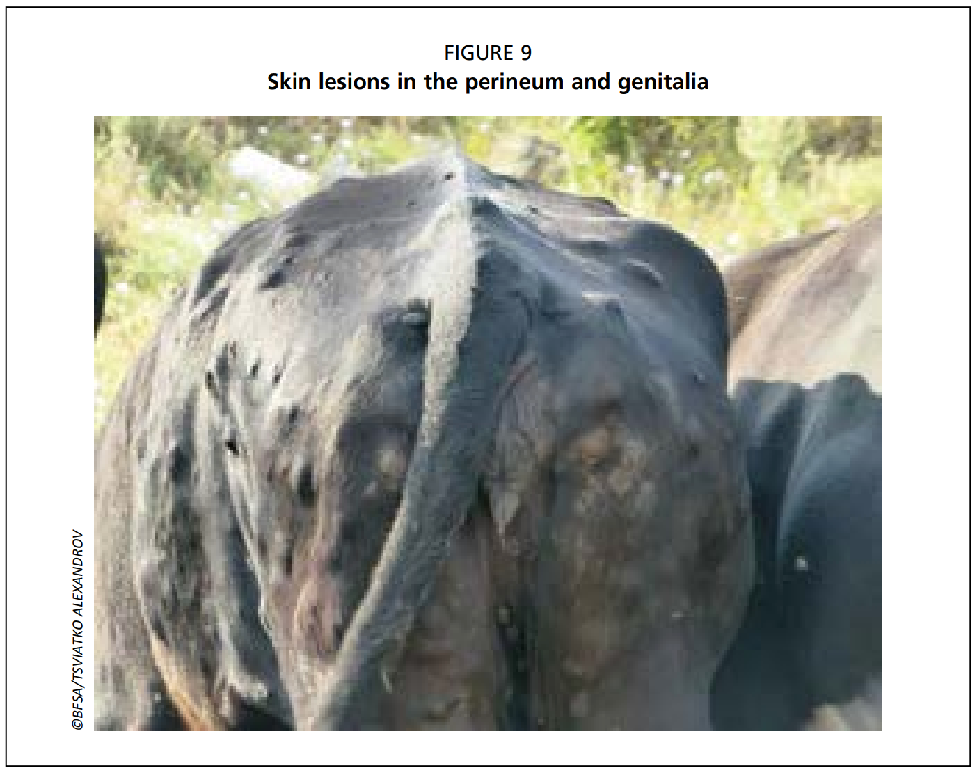
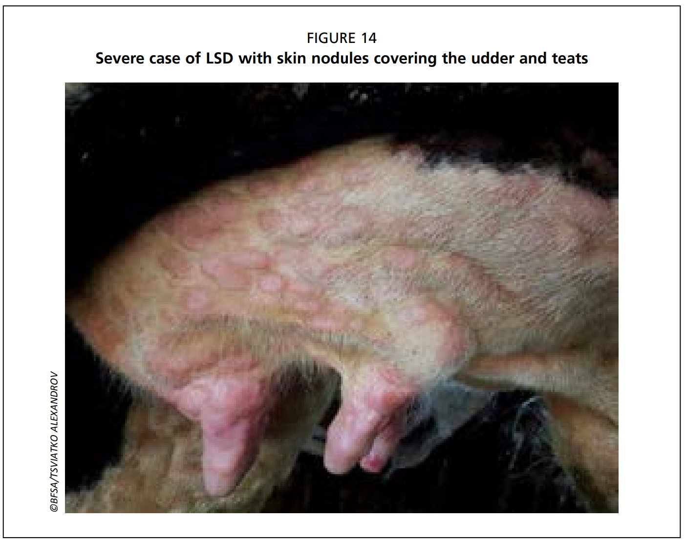
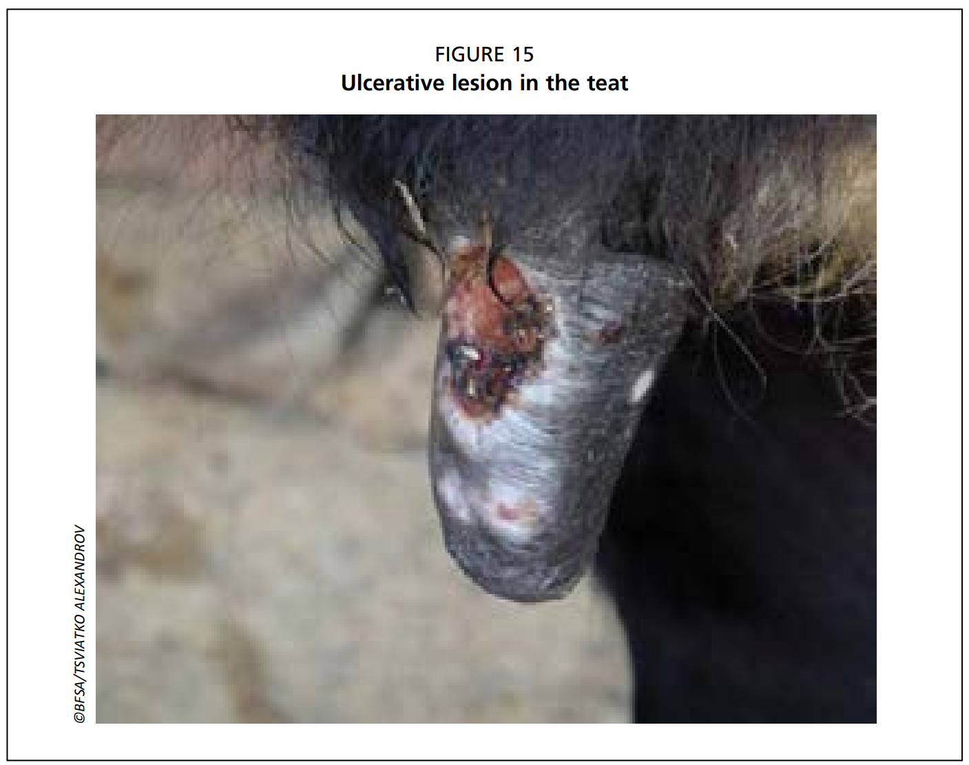
- Predilection sites are the skin of the head, neck, perineum, genitalia (Fig. 9), udder (Figs. 14 & 15) and limbs.
- Deep nodules involve all layers of the skin, subcutaneous tissue and sometimes even the underlying muscles.
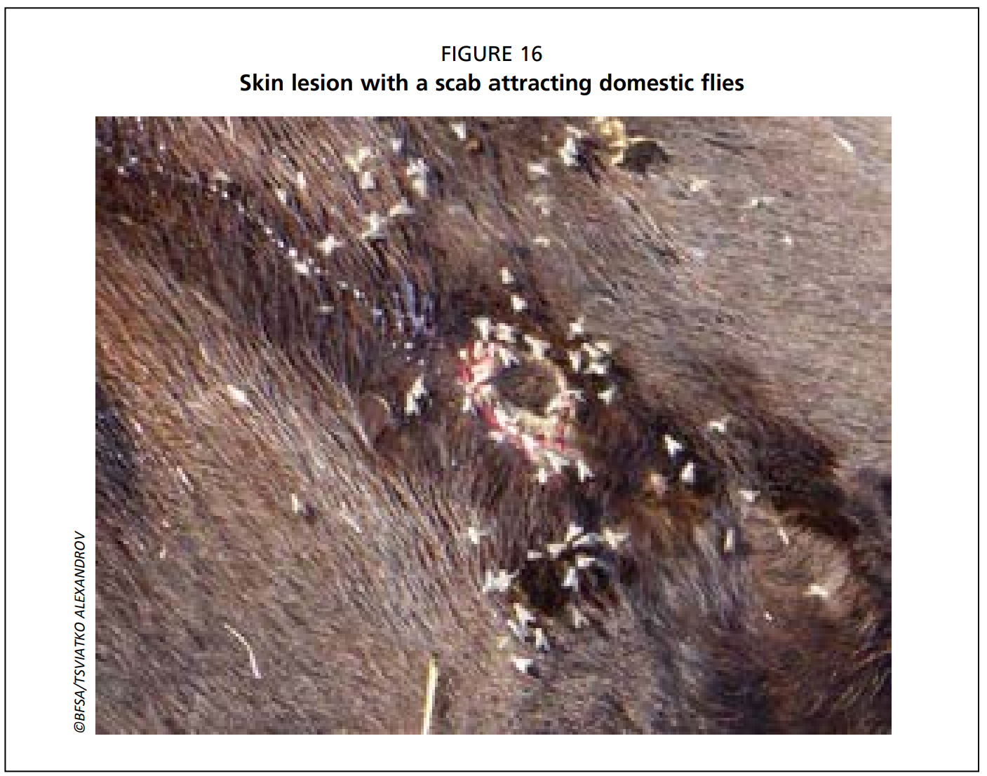
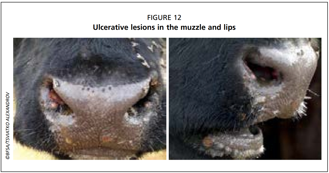
- Necrotic plaques in the mucous membranes of the oral and nasal cavities cause purulent or mucopurulent nasal discharge and excessive salivation, containing high concentrations of virus (Fig. 12).
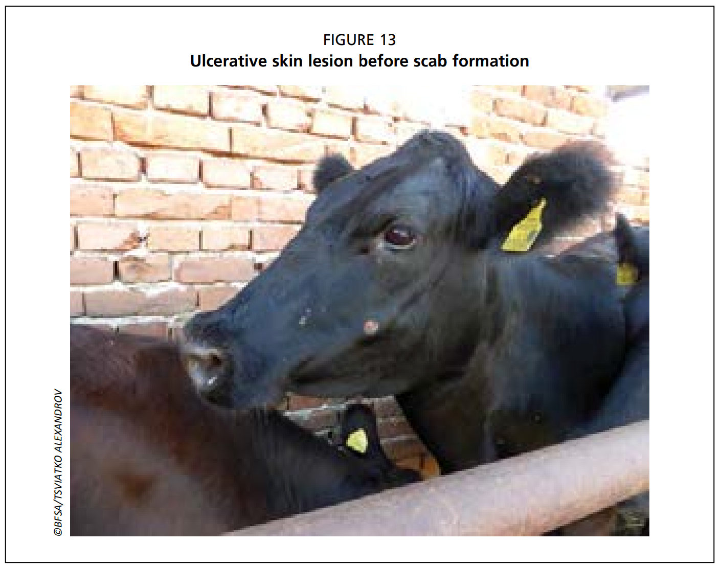
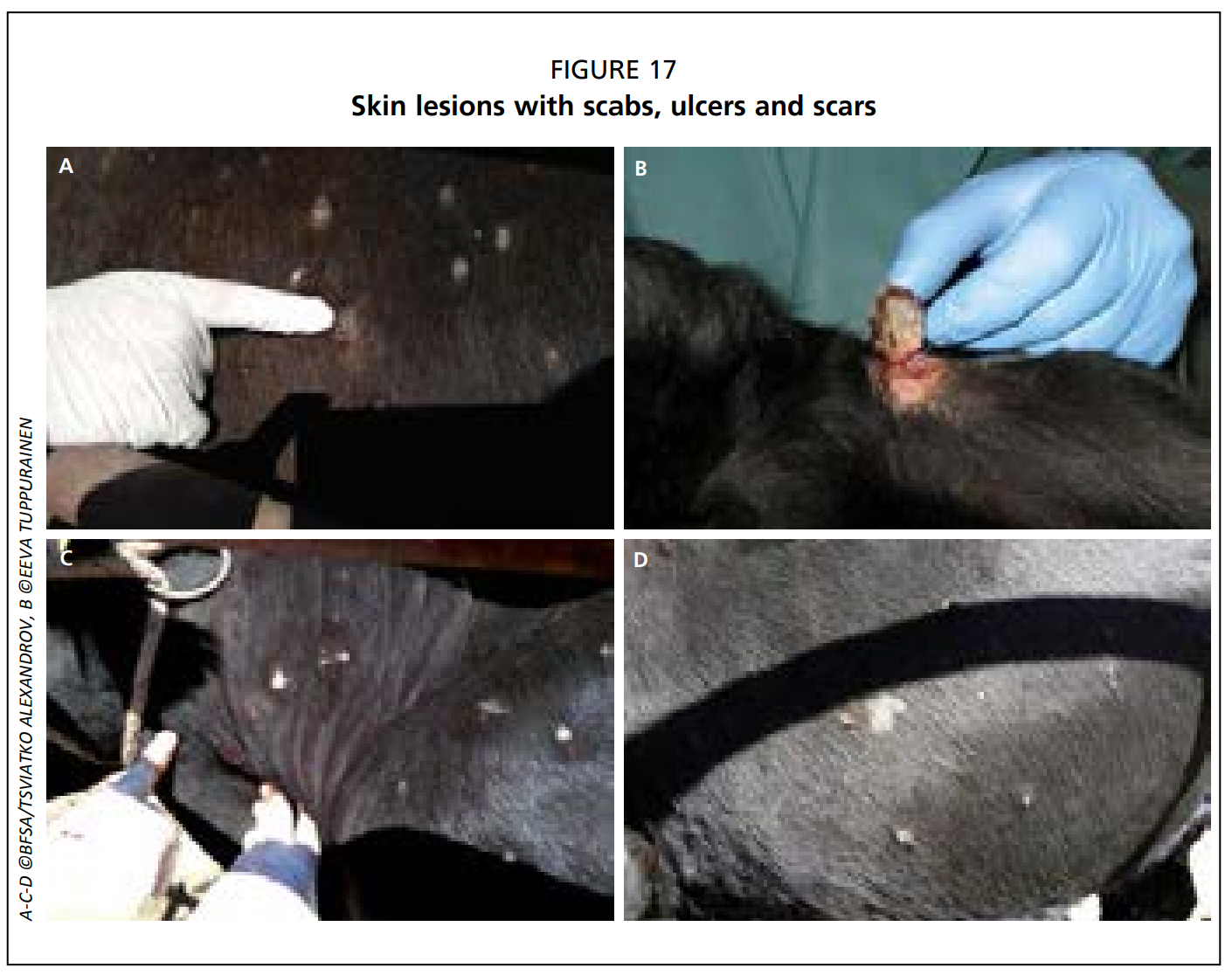
- Typically, the centre of the lesion ulcerates and a scab forms on top (Figs. 13, 16 and 17).
- Skin nodules may persist for several months.
• Sometimes, painful ulcerative lesions develop in the cornea of one or both eyes, leading to blindness in worst cases (Fig. 11).
• Skin lesions in the legs and on top of the joints may lead to deep subcutaneous infections complicated by secondary bacterial infections and lameness.
• Pneumonia caused by the virus itself or secondary bacterial infections, and mastitis are common complications.
• Subclinical infections are common in the field.
When an animal with multiple skin lesions is sent to a slaughterhouse, subcutaneous lesions are clearly visible after the animal is skinned.
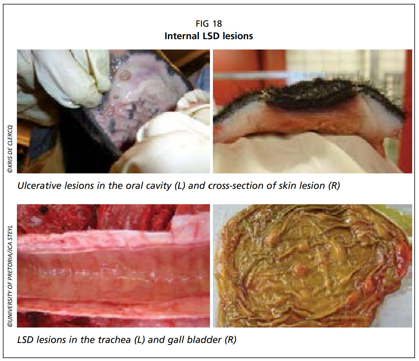
In a postmortem examination, pox lesions can be found throughout the entire digestive and respiratory tracts and on the surface of almost any internal organ (Fig. 18).
Reference:
Tuppurainen, E., Alexandrov, T. & Beltrán-Alcrudo, D. 2017. Lumpy skin disease field manual – A manual for veterinarians. FAO Animal Production and Health Manual No. 20. Rome. Food and Agriculture Organization of the United Nations (FAO). 60 pages.
Headline image courtesy of ©BFSA/Tsviatko Alexandrov