

Understanding the Basics of Mastitis
By G.M. Jones Professor of Dairy Science and Extension Dairy Scientist, Milk Quality & Milking Management, Virginia Tech), T.L. Bailey (Jr., Assistant Professor and Extension Veterinarian, Department of Large Animal Clinical Sciences, Virginia-Maryland College of Veterinary Medicine, Virginia Tech). Table of Contents
Summary
Introduction
Mastitis Causing Bacteria
Effect on Milk Composition
Factors affecting Milk SCC
Cost of Mastitis
References
Summary
Mastitis occurs when the udder becomes inflammed because leukocytes are released into the mammary gland in response to invasion of the teat canal, usually by bacteria. These bacteria multiply and produce toxins that cause injury to milk secreting tissue and various ducts throughout the mammary gland. Elevated leukocytes, or somatic cells, cause a reduction in milk production and alter milk composition. These changes in turn adversely affect quality and quantity of dairy products.Introduction
Before being able to develop or evaluate the effectiveness of a farm's milking management and mastitis prevention and control program, one must have some understanding of what it is that one is attempting to manage or control. According to National Mastitis Council's Current Concepts of Bovine Mastitis, mastitis is an inflammation of the mammary gland in response to injury for the purpose of "destroying or neutralizing the infectious agents and to prepare the way for healing and return to normal function. Inflammation can be caused by many types of injury including infectious agents and their toxins, physical trauma or chemical irritants. In the dairy cow, mastitis is nearly always caused by microorganisms, usually bacteria, that invade the udder, multiply in the milk-producing tissues, and produce toxins that are the immediate cause of injury."The teat end serves as the body's first line of defense against infection. A smooth muscled sphincter, which surrounds the teat canal, functions to keep the teat canal closed, prevent milk from escaping, and prevents bacteria from entering the teat. The cells lining the teat canal produce keratin, a fibrous protein with lipid components (long chain fatty acids) that have bacteriostatic properties. This keratin forms a barrier against bacteria. During milking, bacteria may be present near the opening of the teat canal, either through dirty and wet conditions at the teat end, through teat end lesions or colonization, on contaminated surfaces of milking units (liners or claws), or cow prep procedures. Trauma to the teat renders it more susceptible to bacterial invasion, colonization, and infection because of damage to keratin or mucous membranes lining the teat sinus. The canal of a damaged teat may remain partially open. Conditions which contribute to trauma include: incorrect use of udder washes or cleaning compounds, wet teats, improper mixing or freezing of teat dips, frostbite, failure to prep cows or pre-milking stimulation for milk ejection, overmilking, and insertion of mastitis tubes or teat cannulae. Conditions that are associated with high impact force against the teat end propel bacteria through healthy teat ends. This includes liner slips caused by excessive temporary vacuum losses, low vacuum reserve or level, and abrupt milking unit removal without shutting off vacuum, as well as vacuum fluctuations caused by inefficient vacuum regulation, blocked air vents, restrictions in the short milk tube, poor cluster alignment, or poor liner condition. After milking, the sphincter muscle in the teat canal remains dilated for 1-2 hours and bacteria present during this time can enter the teat canal. Examples would be dirty housing or environment, or failure to use teat dipping properly.
An inflammatory response is initiated when bacteria enter the mammary gland and this is the body's second line of defense. These bacteria multiply and produce toxins, enzymes, and cell-wall components which stimulate the production of numerous mediators of inflammation by inflammatory cells. The magnitude of the inflammatory response may be influenced by the causative pathogen, stage of lactation, age, immune status of the cow, genetics, and nutritional status (Harmon, 1994). Polymorphonuclear neutrophil (PMN) leukocytes and phagocyctes move from bone marrow towards the invading bacteria and are attracted in large numbers by chemical messengers or chemotactic agents from damaged tissues. Masses of PMN may pass between milk producing cells into the lumen of the alveolus, thus increasing the somatic cell count (SCC) as well as damaging secretory cells. Somatic cells consist mainly of PMN or white blood cells.
At the infection site, PMN surround the bacteria and release enzymes which can destroy the organisms. The leukocytes in milk may also release specific substances that attract more leukocytes to the area to fight the infection. Numbers of somatic cells remain in large concentrations after bacteria are eliminated until healing of the gland occurs. Clots formed by the aggregation of leukocytes and blood clotting factors may block small ducts and prevent complete milk removal. Damage to epithelial cells and blockage of small ducts can result in the formation of scar tissue in some cases, with a permanent loss of function of that portion of the gland. In other cases, inflammation may subside, tissue repair may occur, and function may return in that lactation or the subsequent one (Harmon, 1994).
Mastitis Causing Bacteria
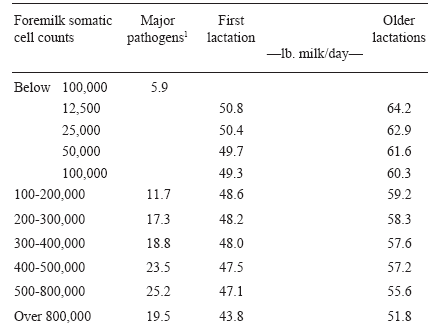
Disease causing bacteria are often referred to as pathogens. The most common mastitis pathogens are found either in the udder (contagious pathogens) or the cow's surroundings (environmental pathogens), such as bedding, manure, soil, etc. Contagious mastitis pathogens (Staphylococcus aureus, Streptococcus agalactiae) are spread from infected udders to "clean" udders during the milking process through contaminated teatcup liners, milkers' hands, paper or cloth towels used to wash or dry more than one cow, and possibly by flies. Although new infections by environmental pathogens (other streptococci such as Str. uberis and Str. dysgalactiae and coliforms such as Escherichia coli and Klebsiella) can occur during milking, primary exposure appears to be between milkings. Coliform infections are usually associated with an unsanitary environment (manure and/or dirty, wet conditions), while Klebsiella are found in sawdust that contains bark or soil. Approximately 70-80% of coliform infections become clinical (abnormal milk, udder swelling, or systemic symptoms that include swollen quarters, watery milk, high fever, depressed appetitie or elevated body temperature). Environmental pathogens are often responsible for most of the clinical cases. About 50% of environmental streptococci infections display clinical symptoms. Sixty to 70% of environmental pathogen infections exist for less than 30 days and are not easily detected. The dry period is the time of greatest susceptibility to new environmental streptococci infections, especially the first 1-2 weeks and the last 7-10 days before calving or early lactation. The incidence at calving is twice what it is at drying off. Infections during the early dry period are controllable by dry cow antibiotic therapy but those in the late dry period are not (Bramley, 1997)1 . In New Zealand, most infections during the dry period and early lactation are due to Str. uberis but S. aureus dominates during lactation. Dry cow therapy will eliminate 70% of environmental streptococcal infections.Subclinical infections are those in which no visible changes occur in the appearance of the milk or the udder, but milk production decreases, bacteria are present in the secretion, and composition is altered. Table 1 describes the negative relationship between SCC and milk yield in 30 Virginia dairy herds. As SCC increased, milk yield was depressed, with the impact greater in older lactation cows than first lactation heifers. Also, a few cows with lower SCC were infected with major mastitis-causing bacteria and the infection rate increased with elevation in SCC. Many of the cows with SCC over 200,000 in Table 1 had subclinical mastitis. At regulatory SCC levels of 750,000, 25% of cows were infected. Even at European regulatory SCC of 400,000, a considerable number of cows in a herd could be expected to be infected.
Bacteria possess a wide array of defense mechanisms in an effort to avoid destruction. Staphylococci produce a toxin that can impede migration of PMN towards chemoattractants. Also, as an infection persists and milk ducts remain clogged, secretory cells revert to non-producing state and alveoli begin to shrink. Substances released by PMN completely destroy the alveolar structure which are replaced by connective and scar tissue. Pockets of infection become walled off and they become difficult to reach with antibiotics. In addition, the clots formed by the aggregation of PMN and blood clotting factors may block small ducts and prevent complete milk removal.
Table 1. Relationship between somatic cell counts, milk production, and intramammary infections.

11% of milk samples from cows within each SCC range with positive culture results for at least one major mastitis pathogen. Jones et al. (1984)
Effect on Milk Composition
Mastitis resulting from major pathogens causes considerable compositional changes in milk (Table 2), including increases in SCC. The types of proteins present change dramatically. Casein, the major milk protein of high nutritional quality, declines and lower quality whey proteins increase which adversely impacts dairy product quality, such as cheese yield, flavor and quality. Serum albumin, immunoglobulins, transferrin, and other serum proteins pass into milk because vascular permeability changes. Lactoferrin, the major antibacterial iron-binding protein in mammary secretions, increases in concentration, likely because of increased output by the mammary tissue and a minor contribution from PMN. Milk protein breakdown can occur in milk from cows with clinical or subclinical mastitis due to presence of proteolytic enzymes. Plasmin increases proteolytic activity by more than 2-fold during mastitis. Plasmin and enzymes derived from somatic cells can cause extensive damage to casein in the udder before milk removal. Deterioration of milk protein as a result of mastitis may continue during processing and storage. Mastitis increases the conductivity of milk and sodium and chloride concentrations are elevated. Potassium, normally the predominant mineral in milk, declines. Because most calcium in milk is associated with casein, the disruption of casein synthesis contributes to lowered calcium in milk.Table 2. Changes in milk constituents associated with high SCC
| Constituent | Normal Milk | Milk with high SCC |
| % | ||
| Fat | 3.5 | 3.2 |
| Lactose | 4.9 | 4.4 |
| Total Protein | 3.61 | 3.56 |
| Total Casein | 2.8 | 2.3 |
| Whey Protein | 0.8 | 1.3 |
| Serum Albumin | 0.02 | 0.07 |
| Lactoferrin | 0.02 | 0.10 |
| Immunoglobins | 0.10 | 0.60 |
| Sodium | 0.057 | 0.105 |
| Chloride | 0.091 | 0.105 |
| Potassium | 0.173 | 0.157 |
| Calsium | 0.12 | 0.04 |
Current Concepts of Bovine Mastitis, National Mastitis Council.
Factors Affecting Milk SCC
The determination of milk SCC is widely used to monitor udder health and, thus, milk quality. SCC are readily available to most dairy farmers through the DHI program. When combined with bacteriological culture results, the factors of greatest importance can be determined. When SCC are elevated, they consist primarily of leukocytes or white blood cells which include macrophages, lymphocytes, and PMN. During inflammation, the major increase in SCC is because of the influx of PMN into milk. At this time, over 90% of the cells may be PMN. Milk from normal (i.e., uninfected) quarters generally contain below 200,000 somatic cells/ml. Many are less than 100,000. One study estimated that 50% of uninfected cows have SCC under 100,000/ml, and 80% have under 200,000. An elevation of SCC (above 300,000 or DHI score 5 and above) is abnormal and an indication of inflammation in the udder. Temporal changes in SCC suggest dramatic changes in the magnitude of the SCC response during the early, acute stages of the infection, reaching a peak SCC within hours or days. Days, weeks, or longer may be required for SCC to decrease after the pathogens have been eliminated. SCC would also be related to number of quarters infected and the amount of milk being produced by each.Age and Stage of Lactation. Milk from uninfected quarters displays little change in SCC as number of lactations or days in milk increase. SCC of milk from uninfected quarters rose from 83,000 at 35 days postpartum to 160,000 by day 285. SCC of milk from quarters infected with S. aureus rose from 234,000 to 1 million over the same period. SCC in uninfected quarters should be less than 300,000 by 5 days postpartum.
Limitations of SCC. The interpretation of SCC records is particularly applicable to herds experiencing infections from contagious pathogens (S. aureus, Str. agalactiae). Because infections by these pathogens tend to be of long duration, new infections in the herd may lead to increased prevalence of infection and are reflected in elevated SCC for bulk tank or herd average SCC scores. Well-managed herds that have controlled mastitis due to contagious pathogens and have higher average milk production can experience problems with increased cases of clinical mastitis caused by environmental pathogens, yet maintain herd average SCC below 300,000. Data collected on 50,085 Finnish heifers from 1983 through 1991 found that, on the average, a greater percentage of heifers were treated in 1991 than in 1983 (27% vs 18%)(Myllys and Rautala, 1995). Intramammary infections by environmental pathogens tend to be shorter than those caused by contagious pathogens. The period of elevated SCC for these cows would be correspondingly shorter as well. The prevalence of infection by environmental pathogens at any point in time also tends to be low (less than 10% of quarters).
The shorter duration of mastitis caused by environmental pathogens makes the diagnosis of the bacterial cause of mastitis difficult in herds with low SCC. Is one quarter persistently infected or do many infections occur repeatedly in different quarters? Sampling and recording of clinical cases and cows who have elevated SCC is needed to accurately describe herds. Accurate treatment records indicate which cows have been treated, when, and in which quarters. In high prevalence herds (number of cases in herd at any one time), rate of new infections may be low but infections are of long duration with accumulation of infected cows. In high incidence herds (new infections), many cows become infected over a given time, but only a few are infected on a given day due to short duration. Milk samples from all clinical quarters and cows with elevated SCC should be collected and frozen for bacteriological culturing.
Cost of Mastitis
Mastitis costs the U.S. dairy industry about $1.7-2 billion annually or 11% of total U.S. milk production. Much of this cost is attributed to reduced milk production, discarded milk, and replacements which are estimated at $102, $24, and $33 per cow per year. The obvious costs for treatment medication, labor, and veterinary services are low, estimated to total $13 per cow. It must be recognized that mastitis cannot be eliminated from a herd. However, the total cost of mastitis in the average herd enrolled in DHI is approximately $171 per cow, which amounts to $18.6 million cost to the Virginia dairy industry annually. If the goal for each herd was to have an average DHI SCC score of 2.0 and no more than 3 cases of clinical mastitis per 100 cows per month, the average herd of 128 cows could increase annual net income by $57 per cow or $7,296. The fundamental principle of mastitis control is that the disease is controlled by either decreasing the exposure of the teat ends to potential pathogens or by increasing resistance of dairy cows to infection (Smith and Hogan, 1997).References in Refereed Scientific Publications:
Harmon, R. J. 1994. Physiology of mastitis and factors affecting somatic cell counts. J. Dairy Sci. 77:2103-2112.
Jones, G. M., R. E. Pearson, G. A. Clabaugh, and C. W. Heald. 1984. Relationships between somatic cell counts and milk production. J. Dairy Sci. 67:1823-1831.
Myllys, V., and H. Rautala. 1995. Characterization of clinical mastitis in primiparous heifers. J. Dairy Sci. 78:538-545.
National Mastitis Council. 1996. Current Concepts of Bovine Mastitis, 4th ed., Arlington, VA.
April 1998


