Diagnosing and Controlling Johne’s Disease in Beef, Dairy Herds
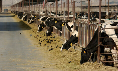
GLOBAL - Johne’s disease, caused by Mycobacterium avium subsp. paratuberculosis (MAP), affects dairy and beef cattle and can cause a range of symptoms including diarrhea, wasting and a dramatic drop in (milk) yield. 27 October 2016
 3 minute read

However, many infected cows remain asymptomatic and pass bacteria onto other cows and their own calves – thereby keeping the infection circulating within the herd.
"If you suspect you have lost animals due to MAP infections or feel that the performance of your herd is insufficient, work with your veterinarian to test the whole herd and determine whether the bacterium is circulating," said Claudia Trummer DVM, PhD, Product Application Specialist at Thermo Fisher Scientific.
"The next step is to decide a plan of action either by reducing the bacterial load by removing the high shedders or eradication, which requires an intensive hygiene management program. There isn’t an easy solution for Johne’s disease, but your veterinarian can help you to determine the best plan for your farming operation."
Diagnosing Johne’s Disease
Only a small proportion of animals will actually develop overt clinical signs and are easily identified and removed from the herd. The identification of subclinical disease in animals, which can shed the organism over long periods and thus be the source of infection for other members of the herd, is crucial for disease control.
"Animals are usually infected at a young age and it can take years until clinical signs appear, but incubation times are highly variable," Dr. Trummer said.
The illustration with the overlapping circles shows that not all cattle are detected with either test.
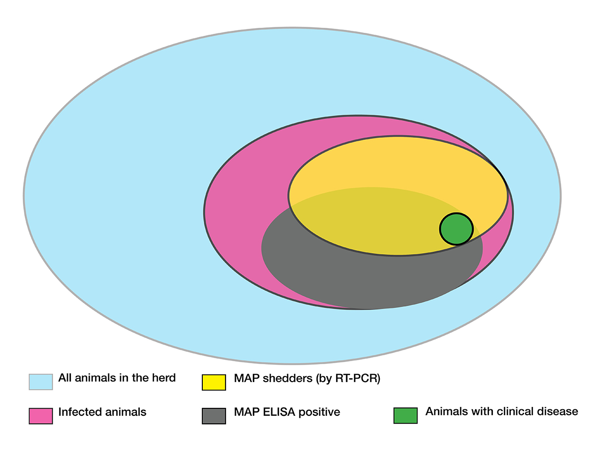
Of the MAP infected animals (pink) in a herd the majority will show an immune response (grey), however, some animals will not develop antibodies.
The majority of animals shed low amounts of bacteria and shedding in these animals occurs intermittently. Only a percentage of infected animals shed higher amounts of MAP (yellow). PCR tests are used to detect shedding animals. These tests are able to detect the pathogen itself (or rather the pathogens’ DNA) with very high sensitivity.
A limitation of the detection with PCR tests is that MAP bacteria are not homogeneously distributed in feces, but rather occur in clusters and can therefore be missed. In addition, PCR also detects transiently infected animals, i.e. animals which have ingested bacteria, but will not develop an infection and do not have to be removed from the herd.
As a rule of thumb, repeated positive PCR tests are indicative for productively infected animals. In these animals the amount of bacteria shed tends to correlate with the appearance of clinical signs.
Because of the different responses of animals to the infection, there is no silver bullet for MAP control programs. Different types of tests are needed to detect MAP infections depending on the stage of disease.
"Using a combination of both ELISA and PCR offers the best solution for comprehensive herd monitoring that can be adapted to your operation’s specific needs – whether that’s testing high risk animals in your herd, individual animals or new additions coming into the herd. With both ELISA and PCR tests, results are available in just a few hours rather than days or weeks as seen with bacterial culture methods," she said.
Thermo Fisher Scientific offers the largest portfolio of diagnostic tests and sample preparation products for MAP. Our diagnostics tools can help farmers deal with this difficult disease and make the best decisions for the herd with confidence.
To learn more about our robust portfolio of PCR and ELISA based MAP diagnostic tests, click here.
TheCattleSite News Desk